Editor’s Note: This post references AcuGraph 5, which has been succeeded by AcuGraph CS. The clinical concepts discussed remain applicable to current practice.
Have you Updated your Software?
This week, we released a new version of AcuGraph 5—version 5.3.0.7. It’s chalk-full of exciting updates and features that we think you’ll really love.

To access the new features, you’ll need to update your software. To do so, open your AcuGraph 5 software. You should automatically be prompted to update the latest version of AcuGraph 5. If no prompt appears, click on the ‘Help’ button at the top of your screen, and select ‘Check for Update’. You’ll be guided through the installation process, and you’ll be ready to start using these new features in a jiffy!
Interested in the other features in this AcuGraph release?
Check out this blog post to read a little bit about the most exciting changes.
If you’re not yet an AcuGraph user and you want to learn more about it, we’d love to get in touch!
You can click here to learn more about AcuGraph and sign up for a free consultation.
Anatomical Reference Images
In this post, we’ll give you an overview of the new anatomical images in the AcuGraph ‘reference’ section. You can read on, or watch the video below to start learning!
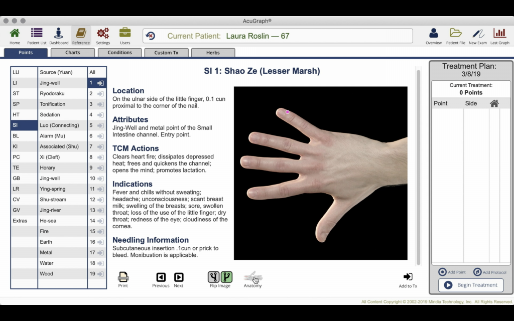
The reference section in AcuGraph has always been a fantastic resource. It includes information such as the location, attributes, TCM actions, indications, needling information and applicable warnings for each acupuncture point.
With this new AcuGraph release, the reference section is even better! It now includes an anatomical reference illustration for each point in addition to the written data. You can access this new feature by clicking on the ‘anatomy’ button below the image of the point.
These anatomical illustrations include not only the location of the point in question, but also the locations of other nearby points and the underlying anatomical structures of the area.
If you want to return to the regular photo view of the point location, you can click the ‘photo‘ icon underneath the anatomical image, and the regular point view will be restored. You can switch back and forth with just one click!

Available Throughout the Software
We wanted to make these anatomical illustrations available in all the places a practitioner might search for an acupuncture point location inside of AcuGraph. So, these images aren’t only available in the reference section—you can find them anywhere in the software where you’d see the point location picture.

For instance, go to your patient list, select a patient and look at his or her last graph. Click on one of the recommended points, and a window will open with reference information and the point location picture.
In the bottom left corner of this window, you’ll find the same ‘anatomy‘ button that we saw in the reference section. Again, you’ll be able to toggle between the two views depending on your needs and preferences.
The same thing happens when you add a point to the patient’s treatment queue and then go look at that queue inside the patient’s file. Once you’ve clicked on the point in the queue, the same window will open, allowing you to toggle between the regular point location photo, and the new anatomical illustrations.
Upgrade to the Latest Software Today!
With these new illustrations, we hope that you will find AcuGraph’s reference section even more intuitive, informative and complete. Make sure not to miss out on this helpful new feature! Update to the latest version of AcuGraph today.
Thanks for Being Part of the AcuGraph Family!
Dr. Adrian Larsen

One Reply to “Anatomical Reference Images – New Acugraph Feature”